One important factor to consider is what file types the software accepts, says John Wiktorowicz, a professor at the University of Texas Medical Branch at Galveston. Some use a proprietary format linked to the company’s imaging system, while many accept generic formats such as JPEG or BMP. Wiktorowicz, director of proteomics for the biomolecular resource facility at the university, prefers to work with TIFFs. “It leaves the values intact,” he explains. Other formats such as JPEG compress the file size and could result in loss of crucial information. “I would never use JPEGs for any quantitative study,” says Wiktorowicz.
Any imaging software should save raw data files, and record any modifications users make to an image. “When it comes time to publish, you need that information,” says Mark Chen, a graduate student at Duke University.

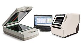
Image Lab™ Software for PC Version 6.0.1. SOFT-LIT-170-9690-ILSPC Image Lab acquisition and analysis software for PC (Windows 7, 32- and 64-bit, and Windows 10, 64-bit), for image acquisition with ChemiDoc XRS+, Gel Doc XR+, Gel Doc EZ, and GS-900 Imaging Systems, and image analysis with all Bio-Rad Imagers. We need Image Lab™ Software #1709690 for bio rad to visualize chemiluminescence, the original software actually got corrupted and getting from company is quite costly any other way to get it? This product is designed to provide high performance blot detection at an affordable price for every lab. Also some info about our TGX Stain-Free FastCCast Acrylamide kits. Best Regards, Sean Taylor Field Application Specialist E: seantaylor@bio-rad.com. For All Technical Support T: 1-800-424-6723 E: techcanada@bio-rad.com. Image Lab software is for personal computers running Windows and Mac OS and is a powerful yet easy to use package for acquisition and analysis of gel and blot images. Image Lab features simplified lane loading normalization and automated detection of lanes and bands with complete report generation. Image Lab standard edition can be downloaded free. LabImage 1D gel analysis is a flexible software solution with strong image analysis algorithms, applicable also for DNA or protein analysis. Due to its workflow-based concept, this application has become a prime example of software usability.
For certain users, such as those who produce pharmaceuticals, a complete data trail is required by the Food and Drug Administration, as laid down in the Code of Federal Regulations Title 21, Part 11 (CFR 21 Part 11). This is particularly important for drug production and quality control, and some clinical labs may want this added layer of record keeping and password security, says Raymond Miller, a product manager at Bio-Rad in Hercules, California.
The simplest choice, Chen says, is to work with the software that comes with your imager. For researchers who want more options, The Scientist profiles five imaging programs.
IMAGEJ
imagej.net/Welcome
The public domain ImageJ software platform, developed at the National Institutes of Health and augmented by various users, is loved by some and hated by others. ImageJ fan Corentin Cras-Méneur, an assistant professor at the University of Michigan Medical School in Ann Arbor, appreciates the flexibility. “It’s one shop for everything,” he says. “With ImageJ I would analyze Western blots, I would do some quantifications of fluorescent microscopy, I would control the microscope . . . anything you can think of.”
For most users, standard ImageJ should be sufficient to analyze bands on a gel or Western, Cras-Méneur says. But for those who want more, many plug-ins are available; coders versed in the Java language can also create their own plug-ins or macros. For example, Cras-Méneur uses a plug-in that analyzes how background signal varies across an image and subtracts it from bands accordingly, instead of assuming the background is uniform.
PROS
Image Lab For Mac
- With a variety of plug-ins available, ImageJ is flexible.
- It supports stacked images, such as pictures collected from a series of different levels using a confocal microscope.
CONS

- 0007ImageJ wasn’t specifically designed for gel or blot analysis, and can be intimidating to new users. While many common questions are answered in online forums, there’s no tech support line to call if you have a specific query. “It’s a lot easier if you have someone around you who has been using it,” says Cras-Méneur.
- Letitia Jones, a postdoc at the University of Rochester Medical Center in New York, disliked the fact that ImageJ typically uses a single box size for all bands on a gel, even if some bands stretched or smiled. She found that, depending on the box drawn by individual users, the results varied quite a bit. “Two people can have totally different conclusions,” says Jones. She prefers Image Studio (see below) because it allows her to customize the box to each band.
- 0007Juan Pablo de Rivero Vaccari, an assistant professor at the University of Miami Miller School of Medicine, says ImageJ’s results didn’t match what he saw with his own eyes—he could tell a band was darker, but the numbers coming out of the software didn’t back him up. He switched to another program, UN-SCAN-IT gel (see below).
IMAGE STUDIO
www.licor.com/bio/products/software/image_studio_lite
SOME, MORE, MOST: All programs discussed in this article can quantify band weight. LI-COR’s Image Studio shown here.COURTESY OF LI-COR BIOSCIENCESThe Image Studio software comes with imaging instruments from LI-COR Biosciences, but any scientist can download the Lite version. It’s the same software, minus the ability to control imagers.
Most users are happy with the Lite version, notes Jeff Harford, senior product marketing manager at LI-COR in Lincoln, Nebraska. However, LI-COR also sells additional, optional features. These include analysis of two-color or in-cell Westerns; a small-animal imaging tool that allows users to draw shapes around objects such as tumors or organs; and a tool to analyze multiwell plates.
PROS
- Chen says the design reminds him of Microsoft Word or Excel, making Image Studio easy to pick up.
- You can sort your files based on parameters such as image date, the type of analysis, or the fluorescent color channels you used. “It’s easy to find old images,” says Chen.
- You can customize the box around each band, to fit bands that stretched or smiled.
CONS
- The software is primarily focused on a few types of analysis, notes Harford. Scientists who want other functions, such as colony counting or microscopy image analysis, could do it with Image Studio but may prefer to look for software dedicated to their needs.
- 0007Jones notes that many tutorials are video format, while she has a harder time finding the written instructions when she wants them. Harford says written quick-start guides and tutorials are available.
IMAGE LAB
www.bio-rad.com/en-us/product/image-lab-software
LINEUP: Bio-Rad’s Image Lab software can automatically define lanes and graph the density of signal across each, as can UN-SCAN-IT gel and CLIQS.COURTESY OF BIO-RADBio-Rad imagers, such as the Gel Doc or ChemiDoc systems, come with the Image Lab software. Researchers can use the software to control the machine and analyze data right on the spot, or transfer the files to a computer for analysis. The newest version of the software features a touchscreen and controls the ChemiDoc Touch.
Image Lab offers numerous features—so many that some users find it overwhelming. One can annotate bands, compare bands to molecular weight standards, and much more. “We’re trying to move away from the concept of just drawing boxes around bands,” says Miller.
PROS
- The software makes it easy to program your imager for your needs, automatically filling in parameters such as the filters necessary for a Western blot or a Ponceau stain.
- One can perform “total protein normalization,” comparing bands of interest to the total protein in each lane, based on labeling such as Ponceau stain. This is more accurate than the common method of labeling housekeeping proteins such as actin to standardize protein load across lanes, says Miller. The result is a ratio of band intensity to total protein in a lane.
- It automatically subtracts the local background around each band, rather than using a single background value.
CONS
- Users can find the abundance of features and lengthy manual intimidating. Chen, who tried Image Lab, recalls, “I felt like somebody needed to train me how to use it.”
- 0007It isn’t very good at automatic lane detection, though Miller says Bio-Rad plans to improve this feature.
- 0007Image Lab mostly uses Bio-Rad’s proprietary .SCN file format, though users can perform some basic analyses with TIFFs, as well as export TIFFs for use in other programs. The company is considering opening it up to more generic formats, says Miller, but adds that users who don’t have Bio-Rad imagers won’t get much benefit from the software.
UN-SCAN-IT GEL
www.silkscientific.com/gel-analysis.htm
SETTING BOUNDARIES: All programs discussed let you fiddle with band edges based on the image and density plots. Shown here: UN-SCAN-IT gel from Silk Software.COURTESY OF JEFFREY SILKUN-SCAN-IT gel was designed to work with any kind of imaging platform—even a regular office scanner or digital camera. It can quantify Westerns, dot blots, gels, and thin-layer chromatography plates. “I like its simplicity,” says user de Rivero Vaccari. “I just need a pixel ‘thing’ and that’s it.” Scientists typically export the data to other programs such as Microsoft Excel or SigmaPlot for analysis and visualization.
UN-SCAN-IT gel also comes bundled with Silk Scientific’s UN-SCAN-IT software, which analyzes hard-copy graphical input—such as a line graph in an old publication or a strip of paper from an analog electrocardiogram—when you don’t have the original data. It digitizes the graph and extrapolates the numbers that likely generated it.
PROS
• 0007It’s easy to use, says de Rivero Vaccari.
• 0007When you draw a box around a band, the software graphically shows the distribution of pixel intensity. It will automatically suggest the borders of the band, but you can adjust the borders of the box based on the graph. “You can use that scientific intuition,” Silk says.
• 0007When two or three bands overlap, the software will help you distinguish them.
• 0007It’s made by a small, focused company. “You get me when you call for customer support,” says company president Silk.
CONS
• 0007UN-SCAN-IT gel lacks some of the fancy features of some other programs; for example, it can’t process 2-D gels.
• De Rivero Vaccari finds it annoying that the software often stretches his original image in the main viewer window, which can make discrete bands look like smears. Silk says the user can choose to stretch the image or maintain the original aspect ratio.
CLIQS
totallab.com/cliqs/
SIZE IT UP: All programs discussed in this article will compute protein size based on a lane with standard markers. Shown here is CLIQS from Totallab.COURTESY OF TOTALLAB
Totallab’s Core Laboratory Image Quantification Software (CLIQS, a combination of two programs formerly known as Phoretix and Totallab Quant) offers a variety of functions to analyze gels, blots, and microplate arrays, and to count colonies. It can automatically detect lanes and bands, calculate molecular weights based on a standard, and subtract background. It can also perform fairly basic measurements of spots on 2-D gels.
Totallab’s family of imaging programs also includes CLIQS 1D Pro, SameSpots, and SpotMap. CLIQS 1D Pro version includes additional features for analysis of one-dimensional gels and plates. For example, it looks for matches between complex band patterns, such as the restriction fragment length polymorphisms used in DNA fingerprinting.
SameSpots and SpotMap are designed for in-depth analyses of 2-D gels. SameSpots allows you to compare multiple gels and match up the spots between them. SpotMap adds the capability to compare those gels to Westerns, and allows more-extensive image editing; you can add or delete spots. It is particularly useful for analysis of residual host cell protein content in biopharmaceutical production.
PROS
- CLIQS is one program with a wide variety of functions that works with any imager.
- 0007It’s easy to extract the numerical data on the bands for statistical analyses, says Denis Wafula, a postdoc at the University of Maryland, College Park, who used the program during his PhD at Florida Agricultural and Mechanical University in Tallahassee.
- You can use total protein normalization or housekeeping proteins to control for protein levels.
CONS
- 0007It can be a bit difficult to figure out the program at first, though it’s easy once you get going, says Wafula.
- With multiple, overlapping programs available from Totallab, it can be difficult to identify the right choice for your lab’s needs, Wafula adds.
| STATS | |||
| Program/Company | Cost | Platform | CFR 21 Part 11 compliance (FDA) |
| ImageJ | Free | Mac/Windows/Linux | No |
| Image Studio LI-COR | Lite version is free. Keys for small-animal, microplate, or multiplex Westerns cost $750 each; the key for in-cell Westerns is $2,175. Some of those features come with certain LI-COR instruments. | Mac/Windows | For those purchasing LI-COR instruments, it is possible to add a custom, CFR 21 Part 11–ready option to the package, but customers are still responsible for ensuring compliance. |
| Image Lab Bio-Rad | Anyone can download the software for free simply by creating a Bio-Rad account. | Mac/Windows | A security license costs $3,705. |
| UN-SCAN-IT gel Silk Software | $445 (perpetual) or $129 (annual); demo version allows you to try it with a couple of your own images. | Mac/Windows | No |
| CLIQS Totallab | GBP850; the company can arrange a trial. | Windows | Yes |
Released:
Allows reading Bio-Rad *.1sc image/analysis files.
Project description
Python Package to read, parse, convert *.1sc files generated by Bio-RadQuantity One software.
Documentation at: http://biorad1sc-reader.readthedocs.io/
An optional dependency: numpy. If present, image operations inbiorad1sc_reader will run 4x to 5x faster.
Also installs the following command-line executables:
- bio1sc2tiff
- converts *.1sc files to *.tif images. (type bio1sc2tiff --help)
- bio1scmeta
- reports all metadata contained in each *.1sc file to a text file.(type bio1scmeta --help)
- bio1scread
- reports details on a *.1sc file’s internal structure.(type bio1scread --help)

Current Status
- “Alpha” quality software
- API subject to change
- No known problems in operation.
- Reading Image Data OK
- Saving Image from file to new TIFF file OK
- Reader.save_img_as_tiff()
- Reader.save_img_as_tiff_sc()
- Reading all metadata OK
- Reader.get_metadata()
- Reader.get_metadata_compact()
- Reading certain image Metadata from Data Block 7 OK
- Reader.get_img_summary()
- Includes:
- Scanner Name: <Scanner Name>
- Number of Pixels: (<img_size_x> x <img_size_y>)
- Image Area: (<img_size_x> mm x <img_size_y> mm)
- Scan Memory Size: <total_img_size> Kb
- Old file name: <old_file_name>
- New file name: <new_file_name>
- <File Directory>
- New Image Acquired
- Save As…
- Quantity One <Quantity One Version String>
- python2 (lack of) support
- I’m currently unwilling to support python2
- If you’d like python2 support, it’s DIY. :)
File Format Documentation
Related Info
- Bio-Rad Image Lab Software
- For Mac, only version 5.x supports 1sc files. (Click on the Download tab to find it.)
- Bio-Rad Quantity One Software
- Generates 1sc files from Bio-Rad instruments.
Release historyRelease notifications | RSS feed
0.7.0
0.6
0.5.1
0.5
0.4
0.3
0.2
0.1.1
0.1
Download files

Download the file for your platform. If you're not sure which to choose, learn more about installing packages.
| Filename, size | File type | Python version | Upload date | Hashes |
|---|---|---|---|---|
| Filename, size biorad1sc_reader-0.7.0-py3-none-any.whl (32.6 kB) | File type Wheel | Python version py3 | Upload date | Hashes |
| Filename, size biorad1sc_reader-0.7.0.tar.gz (30.4 kB) | File type Source | Python version None | Upload date | Hashes |
Hashes for biorad1sc_reader-0.7.0-py3-none-any.whl
| Algorithm | Hash digest |
|---|---|
| SHA256 | 4e4ebad34a3af57ef0880565659d82af994b7c5739c7e6e2c83eaf9393221a99 |
| MD5 | 9ceccf1c5d2f92aec7e39d67dcee23eb |
| BLAKE2-256 | fd0772e4b7e065a2dbad4556c49453f34060c480974da8bf29ae32a44e9ec700 |
Bio Rad Image Software
Hashes for biorad1sc_reader-0.7.0.tar.gz
Bio Rad Image Lab Download
| Algorithm | Hash digest |
|---|---|
| SHA256 | 5d68849c7a6c1d02e4ee7ad4e64bad7da87a42ad08c2c280b55b344503e10fee |
| MD5 | ddf7158a6caa1662c4a3315deecb5ca2 |
| BLAKE2-256 | 1d910034d1e7915347533c952bc4aa828cde303f6e1504cfa134b5f9f3d3d619 |